
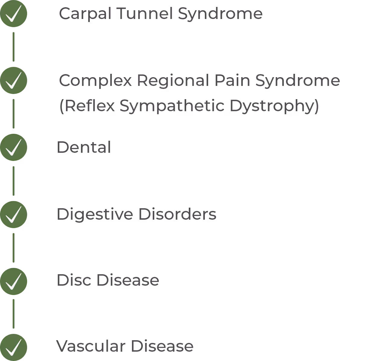
Thermography is one of the best tools for detecting inflammation, which is key because inflammation is often a precursor to disease. Thermography can also identify dysfunction and injury within the body. It does this by taking several scans that provide a temperature map of your body with a digital infrared camera.
At Rapha Health & Wellness, we offer breast scans, as well as full or half body scans for the detection of many conditions. Thermography can assist in diagnosis and treatment as well as help monitor therapy progress. Watch the video to see how thermography screening is beneficial and how it's different than a mammogram.
Schedule your thermography appointment today!
Schedule an Appointment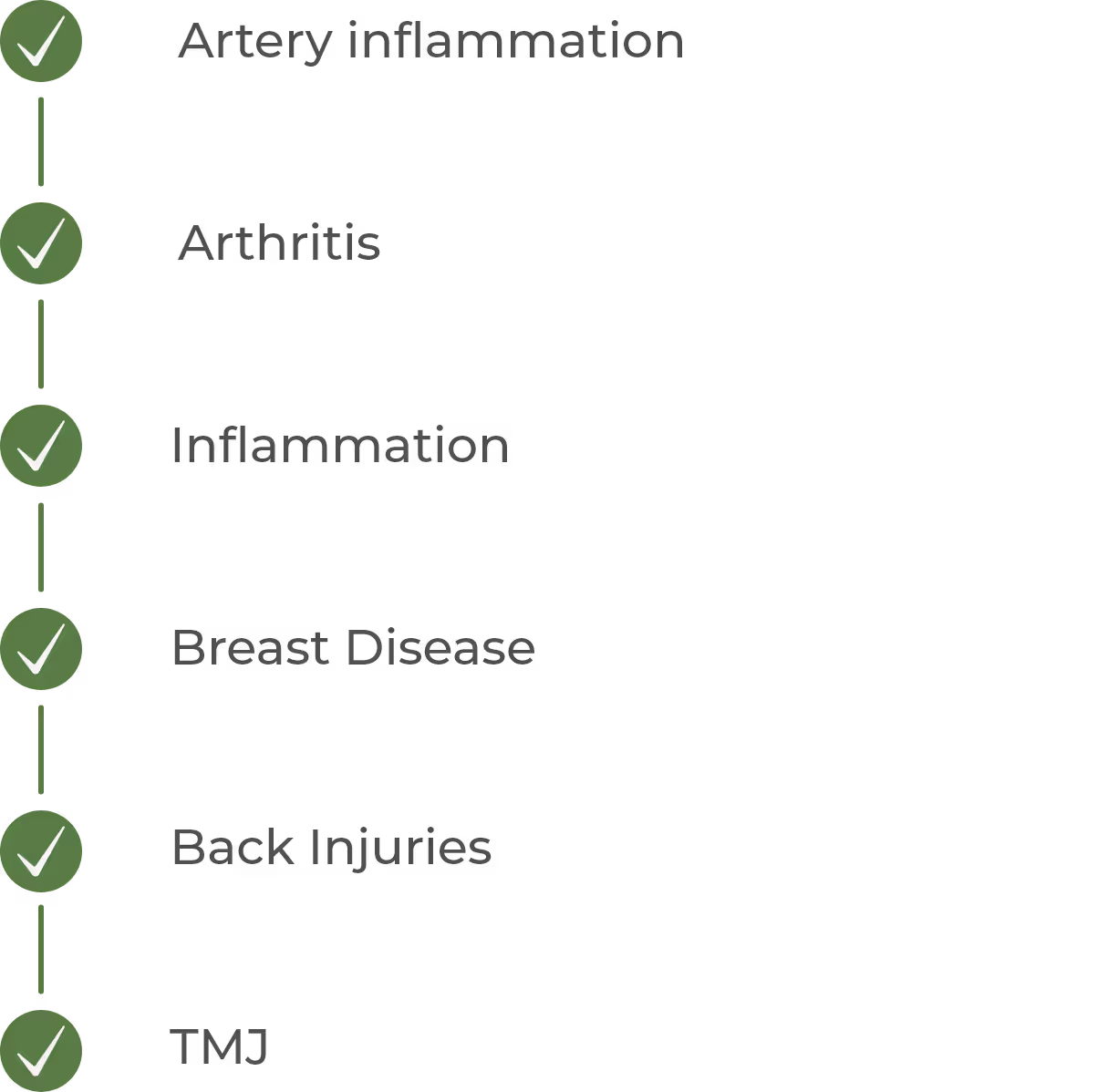
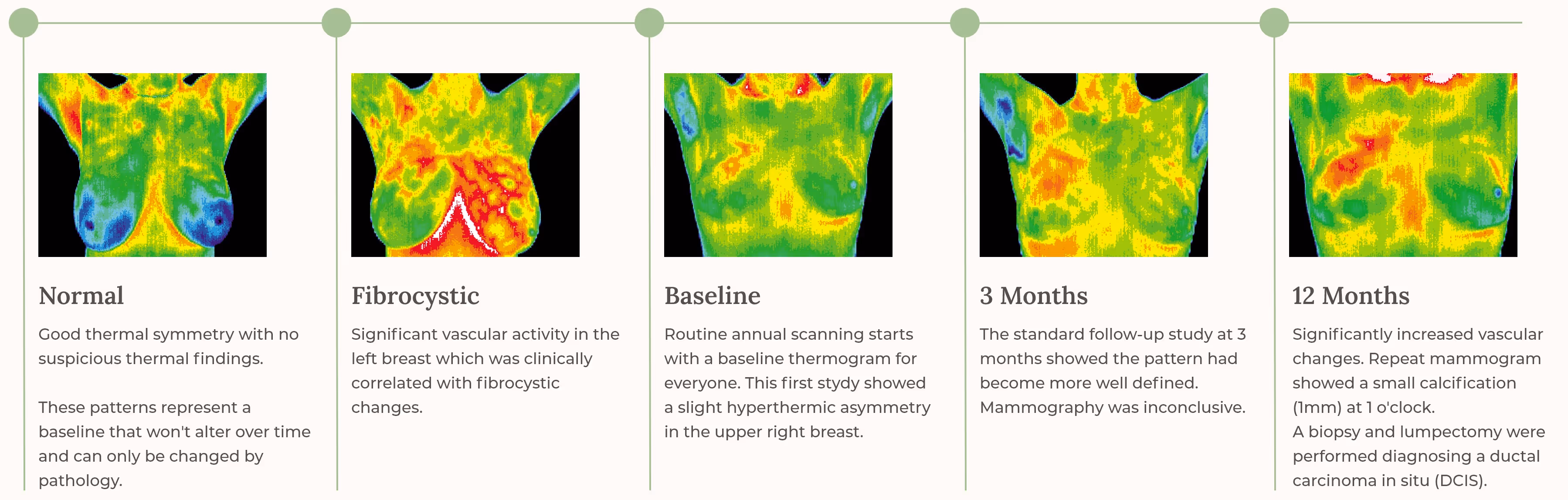
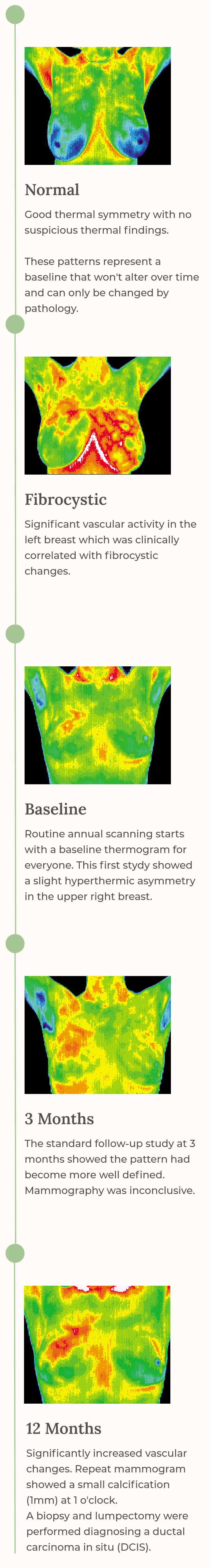
Using Thermography to Detect Abnormal Breast Tissue
Breast thermography is a safe, non-invasive screening tool that detects heat patterns on the breasts using an infrared camera. These patterns are displayed as a color-coded “heat map.” According to the ACCT, changes such as inflammation or increased blood flow can affect skin temperature. Thermography helps identify unusual heat patterns that may need further evaluation. It does not diagnose breast cancer and does not replace mammograms, but provides additional information to monitor breast health overtime. The procedure is painless, radiation-free, and requires no contact or compression, making it comfortable for most women.
Thermography Screening Procedure
A breast thermography baseline is established through two appointments approximately three months apart. This allows the reading physician to determine whether the thermal patterns seen in the first scan remain consistent over time. If the patterns are stable, they create a reliable baseline for comparison with future annual screenings. A physician’s report is typically provided about one week after the second scan, finalizing the baseline and noting any recommendations, such as clinical correlation, if needed.

Medical thermography is not an alternative to mammography, as the two tests serve different purposes. Thermography uses infrared imaging to detect temperature variations that may reflect physiological changes, while mammography uses X-rays to visualize breast structures and identify masses. Since thermography is painless, non-invasive, and radiation-free, it can be safely used for women of all breast types, including those with implants, as part of an overall breast health monitoring program. We also strongly encourage all women to perform monthly self breast exams so they can become familiar with their normal breast tissue and notice changes as early as possible.
Schedule an appointment